

Placental & Perinatal Tissue Biology
Placental and perinatal tissues are specialized fetal structures that develop during pregnancy to support growth, protection, nutrient exchange, and normal fetal development. These tissues—including the placenta, amnion, chorion, umbilical cord, and Wharton's Jelly—perform distinct biological functions while working together as an integrated anatomical system throughout gestation.
Understanding the normal biology of placental and perinatal tissues provides important scientific context for developmental biology, tissue science, extracellular matrix research, and regenerative medicine literature. Their unique anatomy, structural organization, and molecular composition have made these tissues the subject of extensive scientific investigation across multiple areas of biomedical research.
This educational resource explains the normal anatomy, biological organization, and physiological functions of placental and perinatal tissues. It does not discuss specific products, clinical applications, or regulatory classification. Those topics are addressed separately throughout the Regulatory Foundations and Supplier Evaluation Process collections.
What Are Placental & Perinatal Tissues?
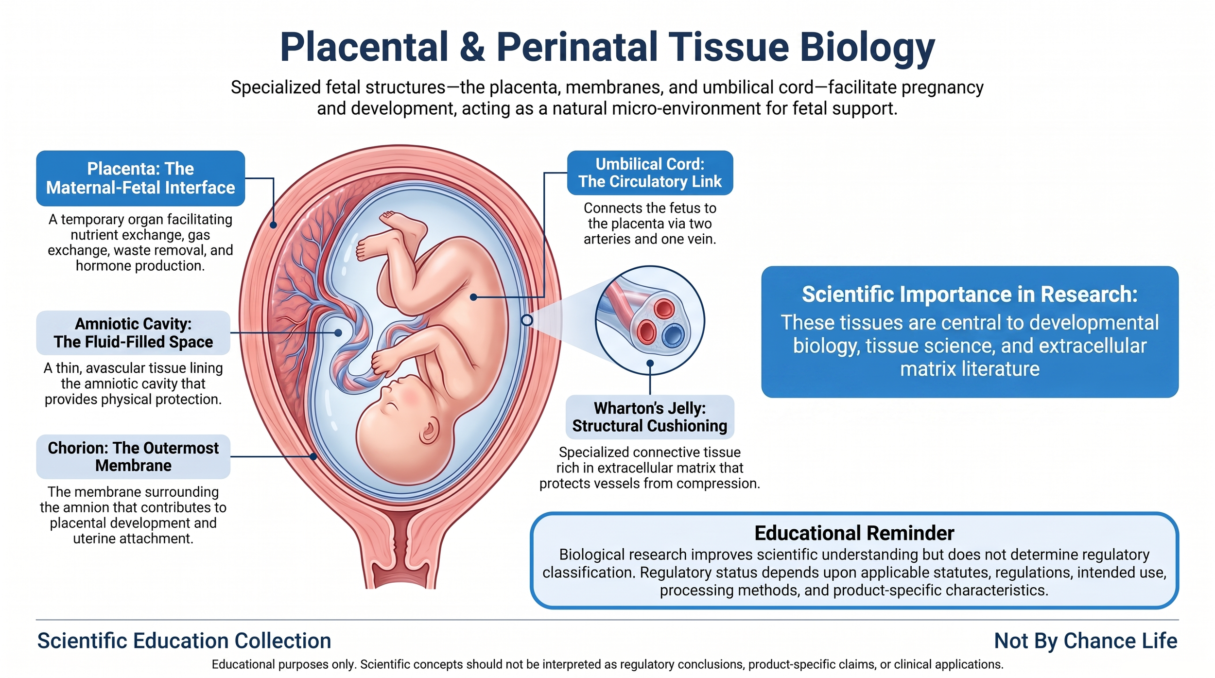
Placental and perinatal tissues are specialized anatomical structures that develop during pregnancy to support fetal growth, protection, nutrient exchange, and normal development. Although each tissue performs distinct biological functions, they operate together as an integrated system that maintains the environment necessary for healthy fetal development throughout gestation.
The term placental tissues generally refers to the placenta and its associated fetal membranes, while perinatal tissues commonly include the placenta, amnion, chorion, umbilical cord, and Wharton's Jelly. These tissues originate during embryonic development and are temporary structures that normally function only throughout pregnancy before being naturally separated following birth.
Each tissue possesses a unique anatomical organization and extracellular matrix composition that reflects its normal biological role. The placenta functions as the interface between maternal and fetal circulation, the amnion and chorion form protective fetal membranes, the umbilical cord provides the vascular connection between the fetus and placenta, and Wharton's Jelly surrounds and protects the umbilical vessels.
Because of their specialized structure and developmental biology, placental and perinatal tissues have become important subjects of scientific investigation within developmental biology, extracellular matrix research, tissue engineering, and regenerative medicine literature. Understanding their normal anatomy provides essential scientific context for interpreting peer-reviewed publications while maintaining a clear distinction between biological science and regulatory evaluation.
Throughout this page, these tissues are discussed solely from the perspective of normal human biology. Scientific descriptions of their structure and function should not be interpreted as evidence of regulatory classification, product characteristics, or clinical application.
The Placenta
The placenta is a temporary fetal organ that develops during pregnancy and serves as the primary interface between maternal and fetal circulation. It is responsible for supporting fetal development by facilitating the exchange of oxygen, nutrients, metabolic waste products, hormones, and other biologically important substances between the mother and fetus while maintaining separate circulatory systems.
Structurally, the placenta consists of highly specialized fetal tissues that attach to the uterine wall and develop an extensive vascular network. This organization allows efficient exchange across the placental barrier while supporting normal fetal growth throughout pregnancy. The placenta also functions as an endocrine organ by producing hormones that contribute to maintaining pregnancy.
Scientific literature frequently examines placental biology because of its unique developmental origin, specialized vascular organization, extracellular matrix composition, and complex interactions between maternal and fetal tissues. These characteristics make the placenta an important model for studying developmental biology, tissue organization, extracellular matrix biology, and fetal physiology.
Although the placenta is widely studied throughout scientific literature, descriptions of placental biology represent observations of normal anatomy and physiology. These biological characteristics alone do not determine the regulatory classification or documented characteristics of any Human Cell, Tissue, or Cellular and Tissue-Based Product (HCT/P).
The Amnion
The amnion is the innermost fetal membrane that surrounds the developing fetus and lines the amniotic cavity. This thin, translucent, and largely avascular membrane encloses the amniotic fluid, creating a protective environment that supports normal fetal movement and development throughout pregnancy.
Histologically, the amnion consists of an epithelial layer, a basement membrane, and multiple connective tissue layers containing extracellular matrix components such as collagen, laminin, fibronectin, proteoglycans, and glycosaminoglycans. These structural elements contribute to the membrane's strength, flexibility, and overall organization while maintaining the integrity of the amniotic sac.
The amnion contributes to fetal protection by helping contain the amniotic fluid that cushions the developing fetus from external mechanical forces. Its specialized extracellular matrix also makes the amnion an important area of investigation within tissue biology, extracellular matrix research, and developmental science.
Because the amnion contains a highly organized extracellular matrix with relatively simple tissue architecture, it has been extensively characterized throughout the scientific literature. Understanding this normal biological organization provides useful context for interpreting publications involving placental tissues and extracellular matrix biology.
The Chorion
The chorion is the outermost fetal membrane that surrounds the amnion and contributes to the formation of the placenta during early pregnancy. Together with the amnion, the chorion forms the fetal membranes that enclose the developing fetus while supporting the structural organization necessary for normal gestation.
During embryonic development, the chorion develops numerous chorionic villi that project toward the maternal uterine tissue. These specialized structures contribute to placental formation and increase the surface area available for maternal-fetal exchange. As pregnancy progresses, portions of the chorion become closely associated with the developing placenta while other regions contribute to the fetal membranes.
Like other perinatal tissues, the chorion contains extracellular matrix components, connective tissue layers, and specialized cellular organization that support its normal anatomical structure. Scientific investigations frequently examine the chorion when studying placental development, fetal membrane biology, extracellular matrix organization, and developmental physiology.
Understanding the normal biology of the chorion helps explain its relationship to the placenta and surrounding fetal membranes while providing important scientific context for developmental biology and placental research discussed throughout the scientific literature.
The Umbilical Cord
The umbilical cord provides the vascular connection between the developing fetus and the placenta. Throughout pregnancy, it serves as the conduit through which fetal blood circulates to and from the placenta, supporting the exchange of oxygen, nutrients, and metabolic waste products while maintaining separate maternal and fetal circulatory systems.
A typical umbilical cord contains three major blood vessels consisting of two umbilical arteries and one umbilical vein. These vessels are surrounded by Wharton's Jelly, a specialized connective tissue that cushions the vessels and helps protect them from compression during normal fetal movement throughout gestation.
The umbilical cord also contains an organized extracellular matrix composed of collagen, proteoglycans, glycosaminoglycans, and other connective tissue components that contribute to its structural integrity and flexibility. These extracellular matrix components have been extensively characterized throughout developmental biology and tissue science research.
Scientific investigations involving the umbilical cord frequently focus on developmental anatomy, vascular biology, extracellular matrix organization, fetal circulation, and connective tissue architecture. These studies improve understanding of normal fetal physiology without establishing regulatory classification or clinical application.
Wharton's Jelly
Wharton's Jelly is a specialized connective tissue that surrounds the umbilical vessels within the umbilical cord. It consists of a highly hydrated extracellular matrix containing collagen fibers, proteoglycans, glycosaminoglycans, and other connective tissue components that provide structural support while helping protect the umbilical vessels throughout pregnancy.
Unlike many connective tissues, Wharton's Jelly contains relatively few organized fibers and a substantial extracellular matrix that contributes to its gel-like consistency. This specialized composition allows the tissue to cushion the umbilical arteries and vein while helping resist mechanical compression as the fetus moves within the amniotic cavity.
The extracellular matrix of Wharton's Jelly has become an important subject of scientific investigation because of its distinctive structural organization and biochemical composition. Researchers frequently examine Wharton's Jelly when studying extracellular matrix biology, connective tissue organization, fetal development, and tissue architecture.
Descriptions of Wharton's Jelly throughout scientific literature are intended to explain its normal biological structure and physiological role during fetal development. These scientific observations should not be interpreted as evidence of regulatory classification, product characteristics, or clinical performance.
How Placental & Perinatal Tissues Function Together
Although the placenta, amnion, chorion, umbilical cord, and Wharton's Jelly each possess unique anatomical characteristics, they function together as an integrated biological system that supports pregnancy and fetal development. Their coordinated organization allows the developing fetus to receive nutrients and oxygen, eliminate metabolic waste, remain physically protected, and continue normal growth within a stable intrauterine environment.
The placenta serves as the interface between maternal and fetal circulation, facilitating the exchange of essential substances while maintaining separate circulatory systems. The umbilical cord connects the fetus to the placenta, allowing blood to travel between these structures through two umbilical arteries and one umbilical vein. Surrounding these vessels, Wharton's Jelly provides structural cushioning that helps protect the vascular network from compression during normal fetal movement.
The amnion forms the innermost fetal membrane surrounding the fetus and encloses the amniotic fluid, while the chorion forms the outer fetal membrane and contributes to placental development. Together, these membranes create a protective environment that supports fetal development while maintaining the structural integrity of the gestational sac throughout pregnancy.
Each tissue also contains specialized extracellular matrix components that contribute to its unique mechanical properties. Variations in collagen organization, proteoglycan composition, glycosaminoglycan content, hydration, and connective tissue architecture allow each structure to fulfill its normal biological role while functioning as part of a coordinated anatomical system.
Understanding how these tissues interact provides important scientific context for interpreting developmental biology, extracellular matrix research, and tissue science literature. Their coordinated organization illustrates how multiple specialized tissues contribute to a single integrated biological function during pregnancy.
Placental & Perinatal Tissue Biology in Scientific Literature
Placental and perinatal tissues have been studied extensively throughout developmental biology, obstetrics, anatomy, extracellular matrix research, pathology, and regenerative medicine. Their unique developmental origin, specialized extracellular matrix composition, and temporary physiological role make these tissues valuable subjects for scientific investigation across numerous biological disciplines.
Researchers study these tissues to better understand fetal development, placental physiology, extracellular matrix organization, tissue remodeling, cellular interactions, vascular development, and connective tissue biology. Scientific publications frequently investigate the molecular composition, structural organization, and developmental processes that contribute to the normal anatomy of placental and perinatal tissues.
Many studies also examine the extracellular matrix components present within these tissues, including collagen, laminin, fibronectin, proteoglycans, and glycosaminoglycans. Understanding these structural molecules helps explain the mechanical characteristics and biological organization observed throughout the placenta, fetal membranes, umbilical cord, and Wharton's Jelly.
As with all areas of scientific investigation, individual studies should be interpreted within the context of their experimental design, methodology, and stated limitations. Laboratory findings, animal models, and other experimental research improve scientific understanding but should not automatically be interpreted as evidence of regulatory status, product characteristics, or clinical application.
Developing a strong understanding of placental and perinatal tissue biology enables readers to more effectively interpret peer-reviewed publications, understand biological terminology, and evaluate scientific discussions involving these specialized fetal tissues.
Connecting Placental & Perinatal Tissue Biology to Documentation Review
Understanding the normal biology of placental and perinatal tissues provides valuable scientific context when reviewing technical documentation, peer-reviewed publications, and educational resources related to Human Cells, Tissues, and Cellular and Tissue-Based Products (HCT/Ps). Knowledge of tissue anatomy, extracellular matrix organization, and normal biological function helps readers recognize scientific terminology commonly encountered throughout documentation without determining regulatory classification.
Documentation may reference structures such as the amnion, chorion, placenta, umbilical cord, Wharton's Jelly, extracellular matrix components, or fetal membranes when describing tissue characteristics. Understanding the normal biology of these tissues enables readers to interpret those descriptions within their proper scientific context while recognizing that biological terminology alone does not establish regulatory status or product characteristics.
Regulatory classification under 21 CFR Part 1271 depends upon applicable statutes, regulations, intended use, processing methods, and documented product characteristics rather than the biological origin or anatomical structure of a tissue alone. Likewise, scientific publications describing placental or perinatal tissue biology should not be interpreted as evidence of regulatory compliance, clinical effectiveness, or FDA authorization.
For this reason, placental and perinatal tissue biology is presented within the Scientific Education collection as one component of a broader educational framework. Readers are encouraged to continue through the Regulatory Foundations and Supplier Evaluation Process collections to better understand how biological science, regulatory requirements, and documentation review complement one another while remaining distinct disciplines.
Continue Your Scientific Education
Placental and perinatal tissue biology represents one component of a broader understanding of developmental biology, tissue science, and extracellular matrix research. Readers interested in expanding their scientific knowledge are encouraged to continue through the remaining Scientific Education collection, where each educational resource examines another foundational area of tissue biology while maintaining a clear distinction between biological concepts, regulatory requirements, and documentation review.
Together, these educational resources provide a structured pathway for understanding the biological principles discussed throughout peer-reviewed scientific literature before exploring how those concepts relate to regulatory evaluation and supplier documentation.
- Extracellular Matrix (ECM) Biology
- Extracellular Vesicle & Exosome Biology
- Secretome Biology
- Tissue Preservation Science
- Cell Viability & Post-Thaw Assessment
- Reading Scientific Literature
- Scientific Glossary
Readers seeking additional educational resources may also continue with the Regulatory Foundations collection or explore the Supplier Evaluation Process collection to better understand how biological science, federal regulations, and documentation review work together while serving distinct educational purposes.
This educational resource is provided for informational purposes only. The biological concepts discussed throughout this page are intended to improve scientific literacy and understanding of placental and perinatal tissue biology. Scientific information should not be interpreted as evidence of regulatory classification, FDA authorization, clinical effectiveness, or product-specific performance. Regulatory determinations for Human Cells, Tissues, and Cellular and Tissue-Based Products (HCT/Ps) should be based upon applicable statutes, regulations, FDA guidance documents, intended use, processing methods, and documented product characteristics.