

Cell Viability & Post-Thaw Assessment
Cell viability assessment is an important area of laboratory science that evaluates the proportion of cells remaining biologically intact under defined experimental conditions. Following cryopreservation and controlled thawing, researchers frequently perform viability testing to better understand cellular integrity, laboratory performance, and biological characteristics observed during scientific investigation. These measurements contribute to biological understanding but represent only one component of a broader scientific evaluation.
This educational resource explains the scientific principles underlying cell viability, post-thaw assessment, commonly used laboratory measurement methods, factors that influence viability results, and the interpretation of biological data. The discussion focuses on established scientific concepts presented throughout the peer-reviewed literature and does not address product-specific performance or clinical applications.
Although viability testing is widely discussed throughout tissue science and regenerative medicine literature, laboratory measurements should not be interpreted independently of the methodology used to obtain them. Scientific interpretation requires consideration of preservation methods, tissue characteristics, analytical techniques, experimental conditions, and biological variability. Understanding these factors helps readers interpret published studies more accurately while distinguishing scientific observations from regulatory evaluation.
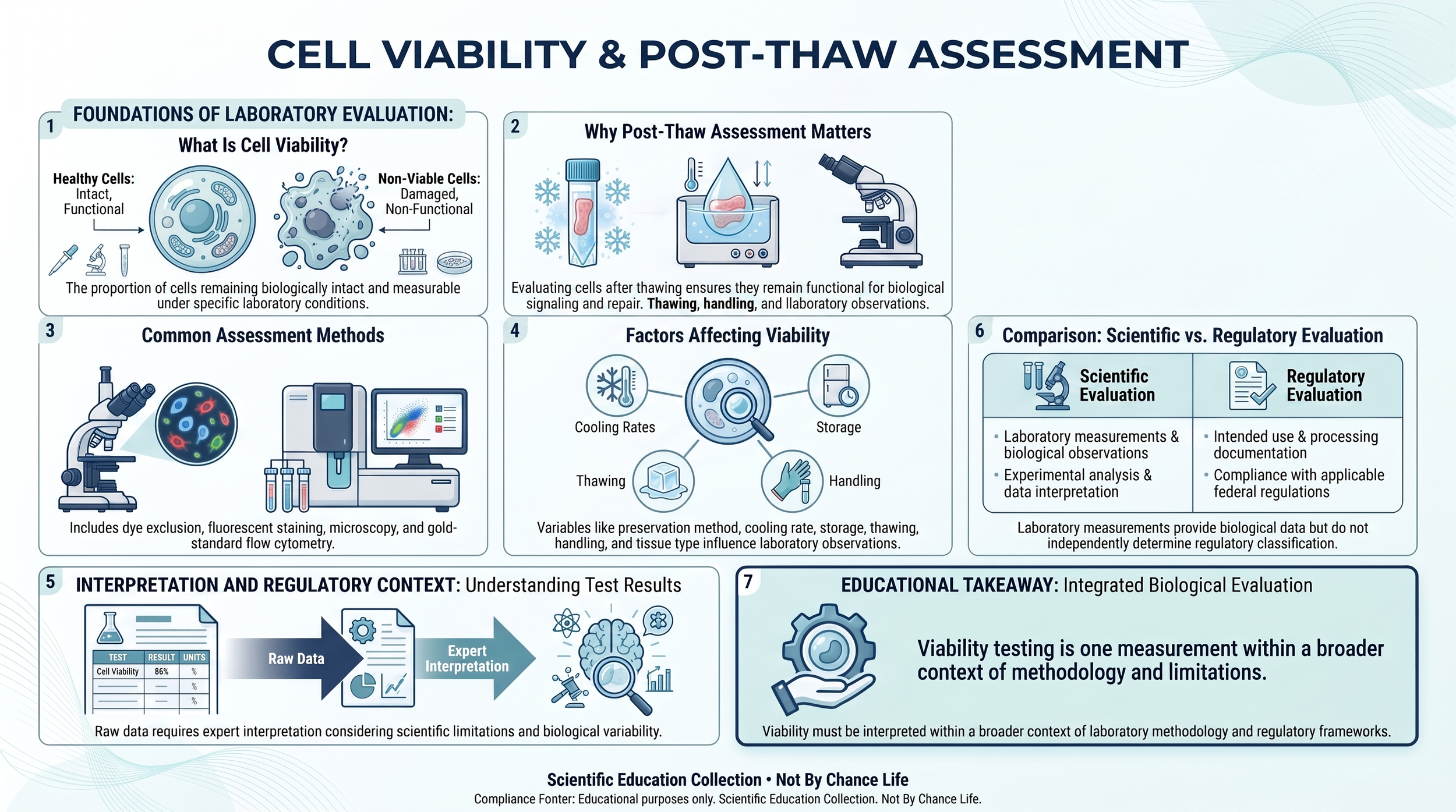
What Is Cell Viability?
Cell viability refers to the proportion of cells that remain biologically intact and measurable under defined laboratory conditions. In scientific research, viability assessments help investigators evaluate cellular integrity following preservation, storage, thawing, or other experimental procedures. Because viability is measured using standardized laboratory techniques, results represent observations obtained under specific testing conditions rather than absolute biological characteristics.
Viability is commonly expressed as a percentage of cells determined to be viable according to the analytical method being used. However, different laboratory techniques evaluate different biological characteristics. Some methods assess membrane integrity, others examine metabolic activity, enzyme function, mitochondrial performance, or fluorescence-based indicators. Consequently, viability percentages obtained using one analytical method may not be directly comparable to results generated using another.
Scientific interpretation therefore requires understanding not only the reported viability value but also the methodology used to generate it. Laboratory protocols, instrument calibration, sample preparation, timing of analysis, operator technique, and statistical methodology may all influence reported measurements. Responsible interpretation considers these variables together rather than relying solely on a single numerical result.
It is also important to recognize that viability represents only one aspect of biological evaluation. Researchers frequently combine viability measurements with structural analysis, extracellular matrix characterization, histological examination, molecular testing, biochemical analysis, and other laboratory techniques to obtain a more comprehensive understanding of preserved tissues.
Understanding the scientific meaning of cell viability helps readers interpret peer-reviewed literature more accurately while appreciating both the value and limitations of laboratory measurements.
Why Post-Thaw Assessment Matters
Following cryopreservation, tissues undergo controlled thawing before laboratory evaluation. Post-thaw assessment allows researchers to examine biological characteristics after tissues have transitioned from frozen storage back to conditions suitable for scientific analysis. Because freezing and thawing involve complex physical and biological processes, post-thaw evaluation provides important information regarding the laboratory characteristics observed after preservation.
Scientific investigations frequently examine how preservation protocols, storage conditions, cryoprotective solutions, cooling rates, thawing procedures, and handling practices influence post-thaw observations. Researchers use these evaluations to better understand preservation biology, optimize laboratory methods, and compare preservation approaches under standardized experimental conditions.
Post-thaw assessment often includes multiple complementary laboratory techniques. In addition to viability testing, investigators may perform microscopic examination, extracellular matrix analysis, molecular characterization, histological evaluation, biochemical assays, and other analytical procedures appropriate to the objectives of the study. Together, these methods provide a broader scientific perspective than viability testing alone.
Because post-thaw observations are influenced by numerous biological and methodological variables, scientific interpretation requires careful consideration of the complete experimental design. Preservation methods, tissue characteristics, laboratory protocols, analytical techniques, and statistical methodology all contribute to the interpretation of reported findings.
Common Laboratory Assessment Methods
Numerous laboratory techniques have been developed to evaluate cell viability following preservation and thawing. Each analytical method measures different biological characteristics and possesses distinct advantages, limitations, and areas of application. For this reason, scientific publications typically identify the specific methodology used when reporting viability results.
One commonly used approach is dye exclusion testing, in which specialized dyes help distinguish cells with intact plasma membranes from those exhibiting membrane disruption. Because membrane integrity represents one indicator of cellular condition, dye exclusion methods provide a rapid and widely recognized laboratory assessment. However, membrane integrity alone does not fully characterize overall biological function.
Fluorescent staining techniques employ fluorescent molecules that interact with specific cellular structures or biological processes. Depending upon the dyes selected, investigators may evaluate membrane integrity, metabolic activity, enzymatic function, mitochondrial activity, or other cellular characteristics. These techniques are frequently combined with fluorescence microscopy or flow cytometry to provide quantitative laboratory observations.
Flow cytometry allows investigators to analyze thousands of individual cells rapidly by measuring light scattering and fluorescent signals as cells pass through specialized instrumentation. This technique enables researchers to examine multiple biological characteristics simultaneously while generating highly detailed quantitative datasets. Flow cytometry has become an important analytical tool across cell biology, immunology, and tissue science research.
Additional laboratory methods—including microscopy, metabolic assays, biochemical analysis, molecular characterization, histological examination, and image-based analysis—may also contribute to comprehensive biological evaluation. Because each technique measures different aspects of cellular biology, researchers often combine several complementary methods to obtain a more complete scientific understanding rather than relying on a single laboratory measurement.
Factors That Influence Viability Measurements
Cell viability measurements are influenced by numerous biological, physical, and methodological variables. Reported laboratory values therefore reflect the combined effects of tissue characteristics, preservation methods, analytical techniques, and experimental conditions rather than representing an isolated biological property. Understanding these influences is essential for accurate interpretation of published research.
Preservation methodology represents one important factor affecting post-thaw observations. Cooling rates, cryoprotective solutions, storage temperature, storage duration, controlled thawing procedures, and handling practices may all influence the biological characteristics observed during laboratory evaluation. Scientific investigations frequently examine these variables individually and collectively to better understand their effects on preserved tissues.
Biological variability also contributes to differences observed between studies. Tissue composition, extracellular matrix organization, cellular density, developmental stage, donor characteristics, hydration, and normal physiological variation may all influence laboratory measurements. Consequently, results obtained from one tissue type or experimental system should not automatically be generalized to other biological settings.
Analytical methodology introduces an additional layer of variation. Differences in laboratory instruments, operator technique, staining procedures, timing of measurements, sample preparation, statistical analysis, and data interpretation may influence reported values even when evaluating similar biological materials. For this reason, responsible interpretation requires careful consideration of both biological and methodological context rather than focusing exclusively on numerical results.
Interpreting Laboratory Results
Laboratory measurements should always be interpreted within the context of the methodology used to generate them. A reported viability percentage represents one scientific observation obtained under defined experimental conditions rather than a complete biological assessment. Meaningful interpretation requires understanding how the sample was prepared, which analytical techniques were employed, when measurements were obtained, and how the resulting data were analyzed.
Researchers typically evaluate laboratory findings alongside additional biological information, including tissue structure, extracellular matrix organization, histological observations, molecular analyses, biochemical measurements, and other complementary data. This integrated approach provides a more comprehensive scientific understanding than any single laboratory value considered independently.
Scientific publications also acknowledge the inherent variability present within biological systems. Differences in tissue characteristics, preservation protocols, laboratory instrumentation, operator technique, and statistical methodology may all contribute to variation between studies. Consequently, responsible interpretation focuses on the broader body of peer-reviewed evidence rather than isolated numerical results reported in individual publications.
Understanding the limitations of laboratory testing is equally important. Every analytical method possesses defined sensitivity, specificity, precision, and methodological constraints. Recognizing these limitations helps readers critically evaluate scientific findings while avoiding conclusions that extend beyond what the available data support.
Developing this analytical perspective strengthens scientific literacy and prepares readers to interpret peer-reviewed publications with greater confidence while maintaining appropriate distinction between laboratory observations, biological interpretation, and regulatory evaluation.
Scientific Evaluation and Regulatory Classification
Scientific laboratory evaluation and regulatory classification serve different purposes and should not be considered interchangeable. Laboratory testing investigates biological characteristics through standardized analytical methods, whereas regulatory evaluation determines whether a Human Cell, Tissue, or Cellular and Tissue-Based Product (HCT/P) satisfies applicable statutory and regulatory requirements based upon documented product characteristics, intended use, manufacturing practices, and supporting documentation.
Measurements of cell viability, post-thaw observations, or other laboratory findings contribute valuable scientific information but do not independently establish regulatory status, FDA authorization, or product-specific performance. Biological measurements represent one component of scientific investigation and should always be interpreted within the broader context of experimental methodology and supporting evidence.
For HCT/Ps regulated under 21 CFR Part 1271, regulatory classification depends upon applicable federal statutes, regulations, FDA guidance documents, intended use, processing methods, and documented product characteristics rather than laboratory viability measurements alone. Scientific observations strengthen biological understanding, whereas regulatory evaluation applies established legal and regulatory criteria.
Maintaining this distinction helps readers evaluate scientific literature responsibly while recognizing that biological measurements and regulatory determinations answer different questions. Scientific investigation improves understanding of tissue biology; regulatory evaluation determines compliance with applicable federal requirements.
Readers interested in the regulatory framework governing HCT/Ps should continue with the Regulatory Foundations collection, while those evaluating supplier information should explore the Supplier Evaluation Process. Together with the Scientific Education collection, these educational resources provide complementary perspectives that support informed interpretation of both biological science and technical documentation.
Related Scientific Education Resources
Cell viability and post-thaw assessment represent one part of a broader scientific understanding of preservation biology, tissue science, and laboratory evaluation. The following educational resources examine related concepts that frequently appear throughout peer-reviewed literature and technical documentation, providing additional context for interpreting biological research and understanding scientific terminology.
- Scientific Education
- Extracellular Matrix (ECM) Biology
- Placental & Perinatal Tissue Biology
- Extracellular Vesicle & Exosome Biology
- Secretome Biology
- Tissue Preservation Science
- Reading Scientific Literature
- Scientific Glossary
Readers seeking additional regulatory context may also benefit from the Regulatory Foundations collection and the Supplier Evaluation Process, which explain how biological science, federal regulations, and documentation review complement one another while serving distinct educational purposes.
This educational resource is provided for scientific education only. Discussions of cell viability, post-thaw assessment, laboratory measurement methods, cryobiology, preservation science, analytical techniques, and related biological concepts are intended solely to improve scientific literacy. They should not be interpreted as evidence of regulatory classification, FDA authorization, clinical effectiveness, or product-specific performance. Regulatory determinations should always be based upon applicable statutes, regulations, FDA guidance documents, intended use, processing methods, and the documented characteristics of the individual Human Cell, Tissue, or Cellular and Tissue-Based Product (HCT/P).